
Have a Question? Need a Customization?
Contact us for device selection, troubleshooting & more!
Response time promise: 1 business day

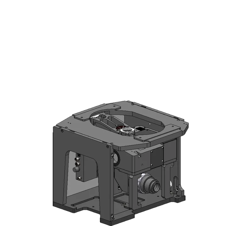
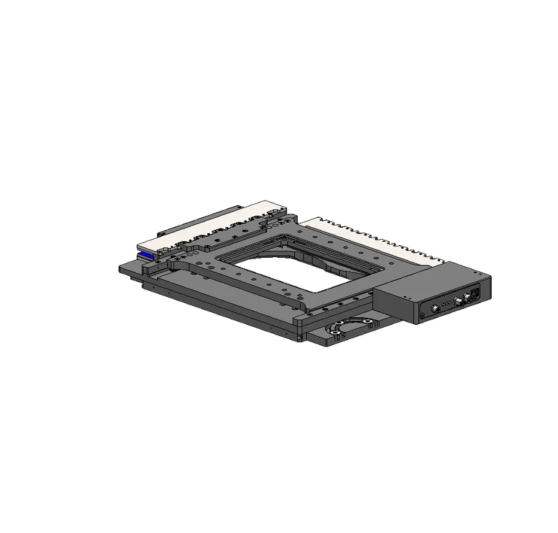

| Stage | Travel Range (mm x mm) |
Repeatability (µm) |
Capacity |
|---|---|---|---|
| X-ADR130B100B | 130 x 100 | 0.5 (precision) | 1 microplate 4 slides 1 petri dish |
| X-ADR250B100B | 250 x 100 | 0.5 (precision) | 2 microplates 8 slides |
| X-ASR100B120B | 100 x 120 | 2 (standard) | 1 microplate 4 slides 1 petri dish |
| X-ASR205B205B | 205 x 205 | 2 (standard) | 2 microplates 1 200 x 200 wafer |
| X-ASR305B305B | 305 x 305 | 2 (standard) | 6 microplates 1 300 x 300 wafer |
Stage travel is typically determined by the size of the sample being imaged. For a single microplate, slide or petri dish, 120 mm x 100 mm travel will be sufficient. For maximum walk-away time, the 305 mm x 305 mm ASR stage can hold up to six standard microplates.
For a complete explanation of specifications impacted by XY stage selection, see our article on XY stage selection.
| Stage | Microplates (qty) | Slides (qty) | Universal |
|---|---|---|---|
| X-ADR130B100B | AM109 (1) | AM242 (4) | AM108 |
| X-ADR250B100B | AM230 (2) | AM231 (8) | ❌ |
| X-ASR100B120B | AM109 (1) | AM242 (4) | AM108 |
| X-ASR205B205B | AM223 (2) | ❌ | ❌ |
| X-ASR305B305B | AM224 (6) | ❌ | ❌ |
| Epi-illuminator | Features |
|---|---|
| MLR3A-T13A | 3-channel (385, 473, 568 nm) |
| MLR3B-T13A | 3-channel (385, white, 625 nm) |
| MLG3A | 3rd party adaptor - 3 mm core liquid light guide - supports field number of 20 mm |
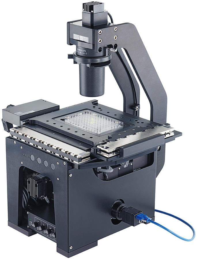
The MVR meets epifluorescence, reflected brightfield, and darkfield imaging needs. The optional MLT transmitted illuminator (pictured here) adds transmitted brightfield and phase contrast imaging capabilities. Third-party light engines are supported via an optional 3 mm liquid light guide adapter.

Contact us for device selection, troubleshooting & more!
Response time promise: 1 business day
| Manual Control | Indexed knob with push switch |
| LED Indicators |  |
| Weight | 3.5 kg (7.716 lb) |
| Compatible Optic Diameter | 25 |
| Number of Optics | 6 |
| Adjacent Optic Change Time | 350 ms |
| Communication Interface | RS-232 |
| Communication Protocol | Zaber ASCII |
| Data Cable Connection | Locking 4-pin M8 |
| Power Supply | 24-48 VDC |
| Power Plug | 2-pin screw terminal |
| Maximum Current Draw | 3300 mA |
| Operating Temperature Range | 0 to 40 °C |
| CE Compliant | |
| Vacuum Compatible |  |

Quickly set up and explore your Zaber devices.
Zaber Launcher is all you need for initial setup, testing and exploration of your Zaber devices. For some simple applications, it's all you'll ever need. It's the one piece of software that we recommend every user of Zaber products should install.
Learn MoreWrite your own code to control Zaber devices.
Ultimately, you will likely want to control your Zaber devices using your own code. We've tried to make that as easy as possible with libraries/APIs that support most popular programming languages and tools.
Whether you need modifications to standard products or have unique stage or system needs, our Product Customization Team is dedicated to building tailored solutions.
Response Time Promise: 1 Business Day
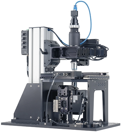
A microscope system to simultaneously image a sample from the top and bottom. Each objective has independent focus control and the upper and lower optical axes are precisely aligned to ensure concentricity. While the solution is a customer configuration, all parts are off-the-shelf products from our wide range of Nucleus microscope modules. The remarkably low cost of $44,000 speaks to the efficiency and affordability of Zaber’s modular approach. Read the case study to learn more.
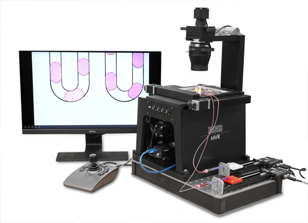
Easily access droplet microfluidics. Zaber devices with integrated controllers and a common Zaber Motion Library API makes it easy to coordinate multiple Zaber stages with the MVR for synchronized flow, imaging, and illumination control. Flow control: Zaber X-LSM100B & X-LSM150A-E03 Illumination: Zaber MLT100AC-T13A Camera: Flir BFS-U3-200S6C-C Objective: Zeiss EC Epiplan NEOFLUAR 2.5x/0.06HD
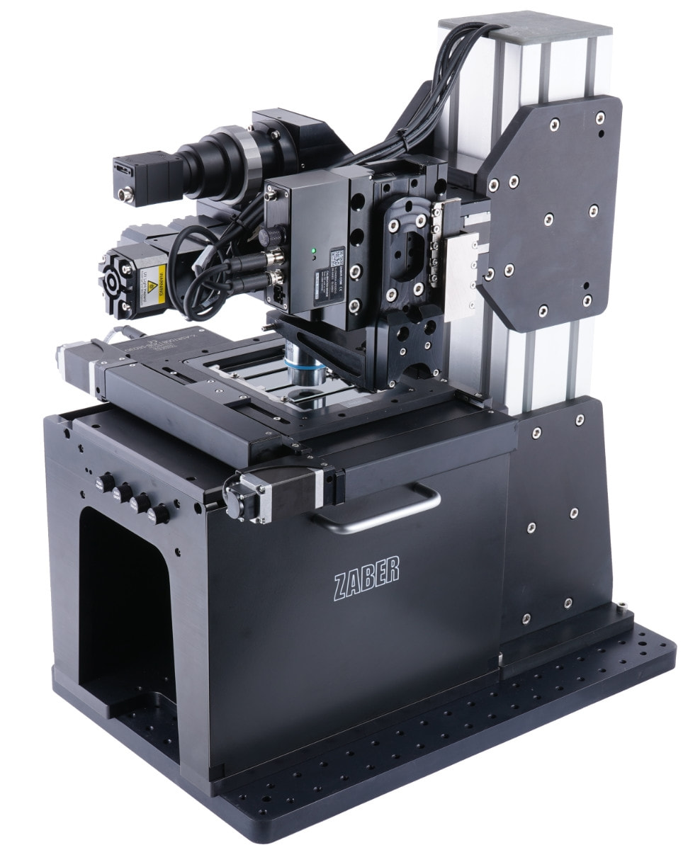
This upright microscope uses Zaber's X-FCR filter cube turret, X-LDA linear stage and X-ASR XY scanning stage to provide upright reflected light brightfield and epi-fluorescence imaging capabilities.
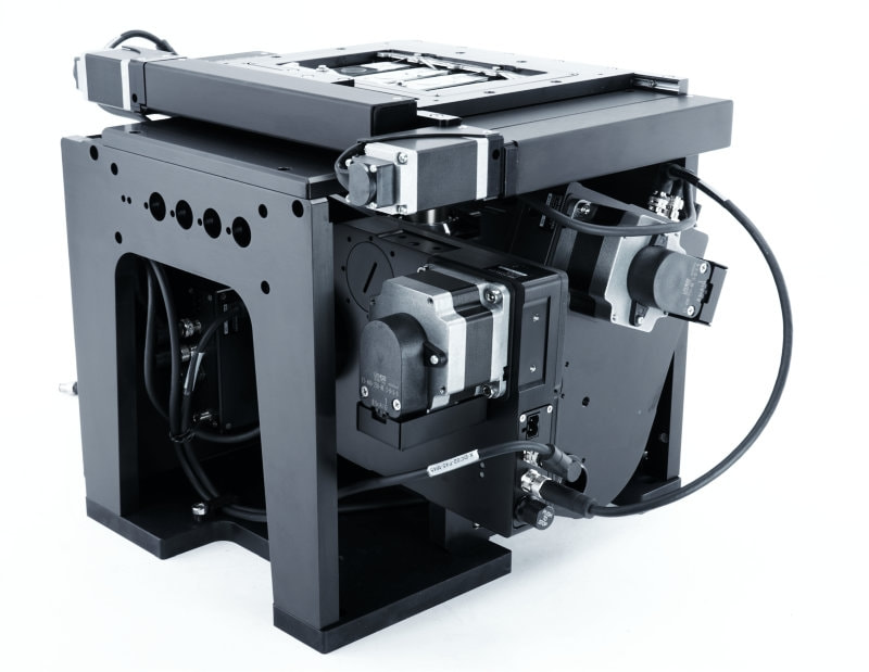
Zaber MVR microscope with a pair of integrated X-FWR filter wheels. This system delivers rapid and individual control of excitation filters, emission filters, and dichroics. The common Zaber motion library and daisy-chained device connections enable the whole system to be controlled using one API and one USB connection. Filter Wheels: Zaber X-FWR06A-E02 Illumination: External Optical system: Zeiss ICS Microscope Stage: Zaber X-ASR100B120B-SE03D12
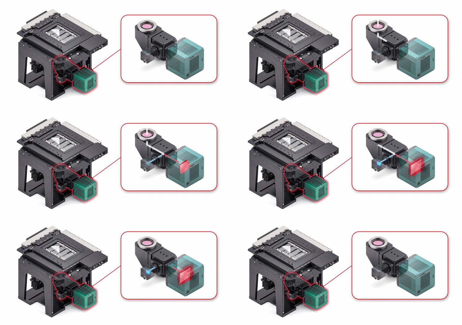
The Challenge: Whether in live cell imaging, semiconductor wafer inspection, or other types of microscopy, microscope users often need to acquire data from two detectors as quickly as possible. Zaber has supported many customers requiring dual camera microscope systems. The three most common reasons are: Higher-Speed: Eliminating mechanical filter switching maximizes imaging throughput. For example, simultaneous imaging of two different fluorophores is essential for investigating rapid processes in live cells. Hyperspectral Imaging: Imaging across an expanded spectral range often requires multiple detectors. The performance of cameras optimized for the visible spectrum falls off beyond 750 nm (Fig. 1). Conversely, cameras which excel well at long wavelengths, do not perform as well in the visible spectrum. A two camera system delivers the best of both worlds. Multi-Mode: Multiple detectors on a single microscope frame can greatly increase a system’s versatility without the need to purchase an entire second microscope. A single system can combine a colour camera for slide scanning with a monochrome camera for fluorescence, or pair a standard camera with more exotic detectors (like SPAD arrays or spectrometers) to easily locate regions of interest. Figure 1. Quantum Efficiency curves of one camera optimized for maximum sensitivity in the visible spectrum (blue curve), and one which trades reduced peak QE for sensitivity over a much larger range of wavelengths (red curve). The Solution: To support two detectors on a single microscope, Zaber created a variant of our MTC tube lens and camera mount module which houses a beam splitter to create two optical paths (Fig. 2). Full compatibility with our Nucleus microscope platform ensures seamless integration into existing automated microscopy workflows. Customers can use a range of different beam splitters including: A 50/50 filter to evenly split the emission wavelengths to both cameras (Fig. 2 A). High or Low pass filters to send specific ranges of wavelengths between the two cameras (Fig. 2 B). A 0/100 filter to transmit or reflect all wavelengths to one or the other camera (Fig. 2 C, D). Figure 2. The optical path can be configured with a beam splitter (A) to send all wavelengths to both paths, or a dichroic mirror (B) to send specific wavelengths to different paths. By inserting or removing a 0/100 mirror (C, D), all light can be directed to one path or the other. Zaber’s dual camera mount supports a range of different cameras and detectors. Options are available for C-mount, F-mount, M42, TFL, SM1, SM2 and Thorlabs cage mount. When pixel-level alignment between the two camera image sensors is required, fine manual adjustment of XY and θ is available. The Engineering Approach: Zaber’s dual camera mount is a drop-in replacement for MTC90 and MTC00 tube lens and camera mount assemblies. This allows existing Nucleus microscope systems to be upgraded. The dual camera mount module uses a DFM1/M Thorlabs 30mm cage cube to house a standard 25 mm x 36 mm beam splitter or dichroic. The optic can be manually moved in and out of the optical path.
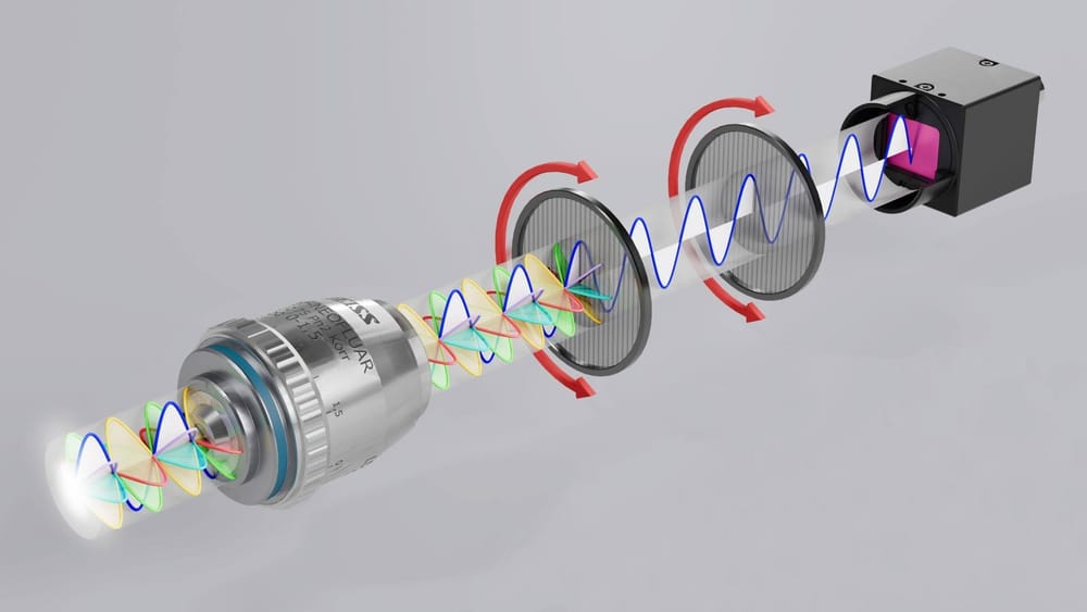
The Challenge: Sensing the degree and angle of linearly polarized light across a sample provides material scientists with a fast, non-contact means of material characterization. Detecting grain boundaries in metals, identifying microplastics in biological materials, and visualizing internal stresses in materials are all enabled by sensing polarized light. Capturing high-resolution polarimetry data across large sample areas requires automation, yet off-the-shelf microscopes with integrated motorized polarizing filters remain scarce. The Solution: A tailored solution delivering a fully motorized, high-resolution polarimetry system based on a Nucleus inverted automated microscope with a pair Zaber motorized rotary stages integrated into the optical path. Sample positioning, polarising filter rotation and image acquisition can be controlled though our free Zaber launcher application. Advanced automation is achieved using the Zaber Motion Library API, which is available for Python, C++, C#, Labview, MATLAB and more. Figure 1. A Zaber Nucleus inverted microscope with motorized polarizing filters mounted between the objective and the tube lens and between the tube lens and the camera mount. The rotation of the polarizing filters can be independently controlled. The Engineering Approach: The low profile and open aperture of Zaber’s X-RSW60A rotary stages allows them to be inserted into the optical path without altering any of the key optical distances for the infinity space or between the tube lens and the image sensor plane. Special care was taken to minimize internal reflections in the optical path when integrating these rotary stages. Optical alignment is critical to imaging performance so custom features were included in the design to enable fine tuning the positioning of each rotary axis during final assembly of the microscope system. Zaber RSW60 rotary stages support standard 25 mm filters. Their position in the optical path ensures the 22 mm maximum field number of the Nucleus microscope is preserved. A large field of view coupled with high speed stages and polarizing filter rotation maximizes data acquisition speed. The fine angular resolution of the X-RSW60A stage supports high resolution polarimetry measurements using high extinction ratio filters. Additional hardware like cameras can be controlled with the Zaber Motion Library API via TTL triggering, minimizing the time, and number of dependencies required to write custom automated polarimetry scanning scripts.