Affordable Dual-Camera Integration for Automated Microscopy
Published on Jun. 26, 2026
The Challenge:
Whether in live cell imaging, semiconductor wafer inspection, or other types of microscopy, microscope users often need to acquire data from two detectors as quickly as possible. Zaber has supported many customers requiring dual camera microscope systems. The three most common reasons are:
- Higher-Speed: Eliminating mechanical filter switching maximizes imaging throughput. For example, simultaneous imaging of two different fluorophores is essential for investigating rapid processes in live cells.
- Hyperspectral Imaging: Imaging across an expanded spectral range often requires multiple detectors. The performance of cameras optimized for the visible spectrum falls off beyond 750 nm (Fig. 1). Conversely, cameras which excel well at long wavelengths, do not perform as well in the visible spectrum. A two camera system delivers the best of both worlds.
- Multi-Mode: Multiple detectors on a single microscope frame can greatly increase a system’s versatility without the need to purchase an entire second microscope. A single system can combine a colour camera for slide scanning with a monochrome camera for fluorescence, or pair a standard camera with more exotic detectors (like SPAD arrays or spectrometers) to easily locate regions of interest.
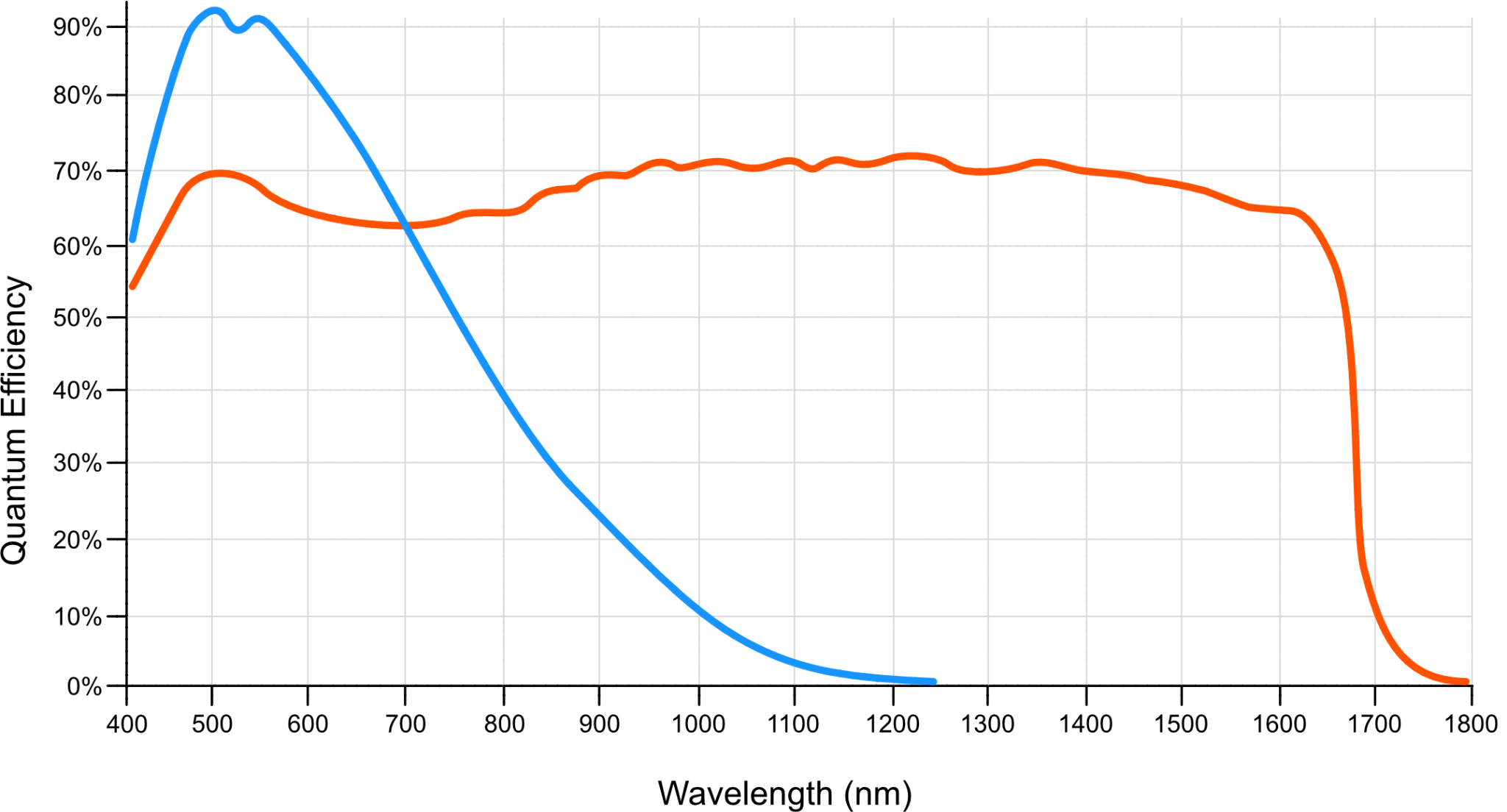
Figure 1. Quantum Efficiency curves of one camera optimized for maximum sensitivity in the visible spectrum (blue curve), and one which trades reduced peak QE for sensitivity over a much larger range of wavelengths (red curve).
The Solution:
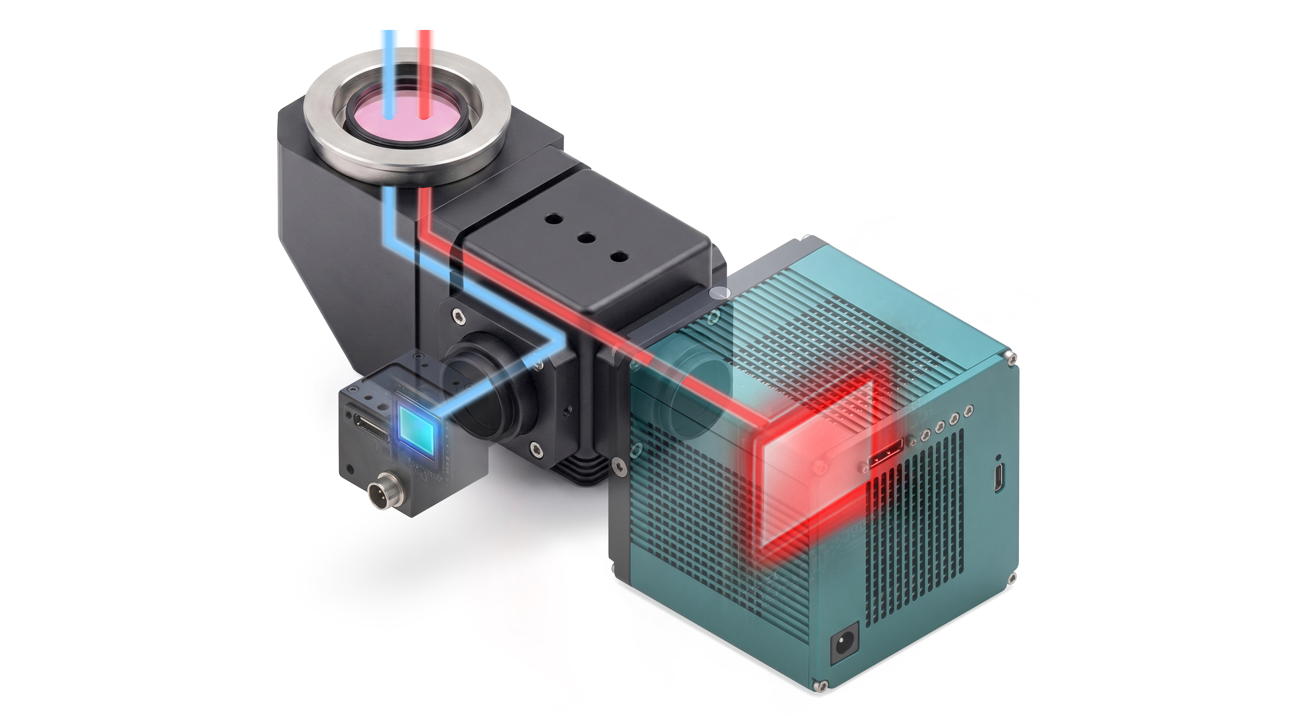
To support two detectors on a single microscope, Zaber created a variant of our MTC tube lens and camera mount module which houses a beam splitter to create two optical paths (Fig. 2). Full compatibility with our Nucleus microscope platform ensures seamless integration into existing automated microscopy workflows. Customers can use a range of different beam splitters including:
- A 50/50 filter to evenly split the emission wavelengths to both cameras (Fig. 2 A).
- High or Low pass filters to send specific ranges of wavelengths between the two cameras (Fig. 2 B).
- A 0/100 filter to transmit or reflect all wavelengths to one or the other camera (Fig. 2 C, D).
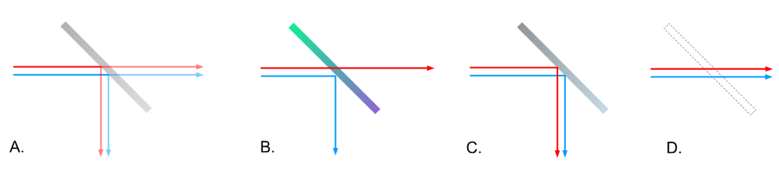
Figure 2. The optical path can be configured with a beam splitter (A) to send all wavelengths to both paths, or a dichroic mirror (B) to send specific wavelengths to different paths. By inserting or removing a 0/100 mirror (C, D), all light can be directed to one path or the other.
Zaber’s dual camera mount supports a range of different cameras and detectors. Options are available for C-mount, F-mount, M42, TFL, SM1, SM2 and Thorlabs cage mount. When pixel-level alignment between the two camera image sensors is required, fine manual adjustment of XY and ፀ is available.
The Engineering Approach:
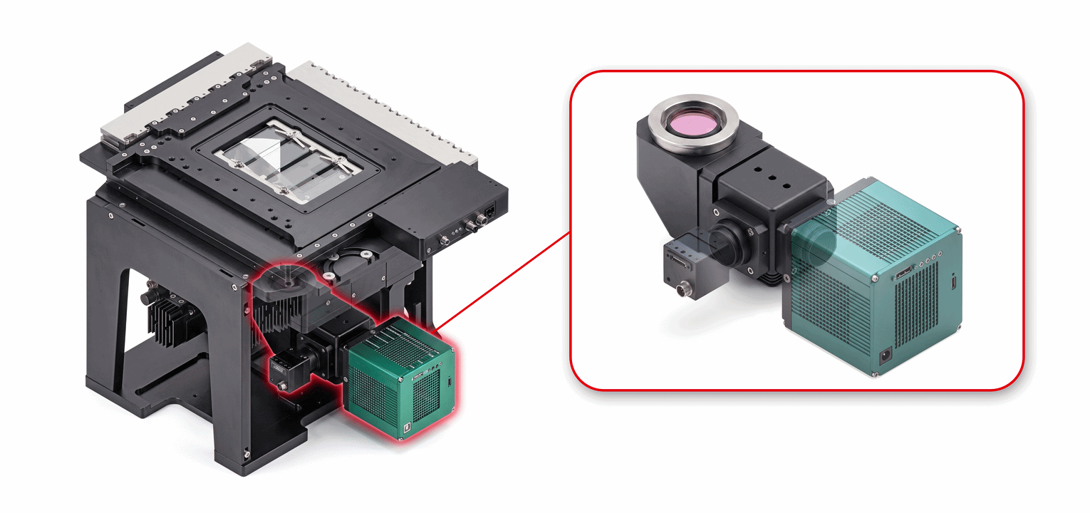
Zaber’s dual camera mount is a drop-in replacement for MTC90 and MTC00 tube lens and camera mount assemblies. This allows existing Nucleus microscope systems to be upgraded. The dual camera mount module uses a DFM1/M Thorlabs 30mm cage cube to house a standard 25 mm x 36 mm beam splitter or dichroic. The optic can be manually moved in and out of the optical path.