Confocal & SIM Imaging: How to Easily Assemble a High-Performance, Affordable System
By Mike Fussell, Life Sciences Product Manager
Published on Nov. 24, 2025
To achieve the spatial resolution and optical sectioning required for many life science research applications, techniques like confocal or structured illumination (SIM) are essential.
Building a spinning disc confocal or structured illumination microscope system requires successful integration of the microscope frame with the confocal or SIM modules at the mechanical, optical, and control levels.
The Zaber Nucleus® microscope platform makes it possible to build advanced imaging systems which are both high performance and affordable.
Meeting the Demands for High-Resolution Imaging
Delivering High-Fidelity Optical Performance
To get the most from a confocal or SIM unit requires high quality objectives and a microscope with an optical path capable of getting the most from them. Nikon, Zeiss and Olympus/Evident all manufacture excellent dry and immersion high magnifications, high numerical apertures objectives offering high field flatness and correction across a wide range of wavelengths. Nexcope’s Nikon compatible objectives are also worth considering. While their best objectives aren’t quite as good as Nikons, they are significantly less expensive.
Ensuring Precise Focus and XY Sample Positioning Repeatability
One design challenge is that high magnification and high numerical aperture objectives have an extremely narrow depth of field. This means, highly repeatable focus with a small minimum step size is necessary when using these objectives.
High magnification objectives also demand highly repeatable XY sample positioning. A highly repeatable XY stage which is capable of consistently returning to the sample position many times can enable an entire tiled array to be captured in one wavelength before switching wavelengths for subsequent XY arrays. This is much faster than imaging each individual XY position at multiple wavelengths before moving on to the next one. A highly repeatable XY stage will ensure the data from each wavelength are precisely co-registered.
Both piezo stages and direct drive linear motors can provide the required level of performance to support these objectives.
- Piezo stages are capable of smaller (0.5 - 2 nm) minimum incremental movements with faster move and settle times, but are more expensive and require large external drivers.
- Linear motor stages have a larger (5 - 20 nm) minimum incremental movement with slower move and settle times, but are much more compact and much less expensive.
Nucleus microscopes with direct drive linear motor focus and XY stages deliver the required performance to ensure your multi-channel XY tiles are precisely co-registered and you can consistently achieve sharp focus, even when working with thin optical sections.
Depending on the optical system of the objectives you wish to use, your Zaber microscope will include an original Zeiss, Nikon or Olympus (Evident) tube lens. Combined with precise machining to ensure the current distances and alignment, Nucleus microscopes deliver the optical performance you expect from your objectives. As a purely digital microscope without additional optics for eyepieces, the optical path is extremely efficient, helping you to reliably detect low intensity signals.
A Modular Platform for Easy Mechanical Integration
Out-of-the-Box Compatibility
Zaber microscopes support integration with confocal scanners from Crest Optics, Yokogawa, and Confocal.nl, and structured illumination units including the VTiSIM (Fig. 1), and Crest optics DeepSIM via the camera mount. The camera mount uses the standard C-Mount. FRAP units from GATACA Systems and Visitron are also supported via coupling to the illumination port.
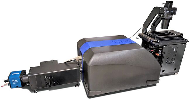
Figure 1. Nucleus microscope coupled to a Visitech VTiSIM
The modularity of the Nucleus platform makes it easy to design a system which meets the needs of your research and your budget. Depending on the imaging modes required, Nucleus microscope systems can be configured with a motorized six position filtercube turret and an integrated 3 channel LED epi illuminator and transmitted illuminator, or with no integrated illumination and a single fixed filter cube.
Flexible Setup for Any Lab Space
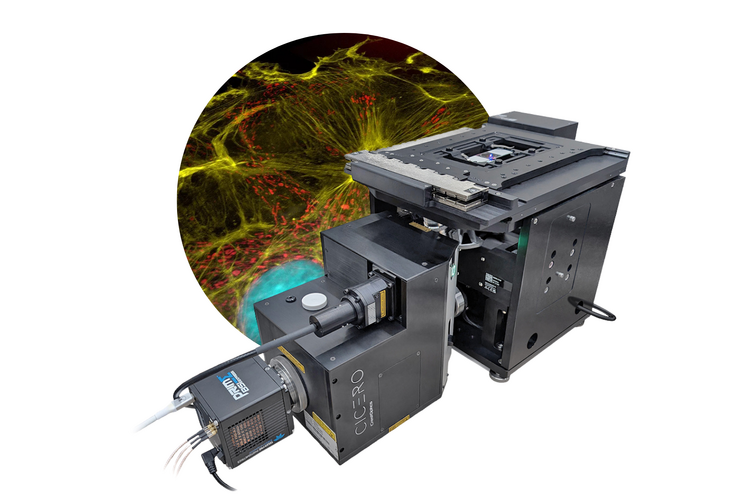
The compact size of the Zaber microscope keeps the system footprint to a minimum. A system combining the Crest Cicero confocal and Nucleus inverted microscope is quite compact (Fig. 2).
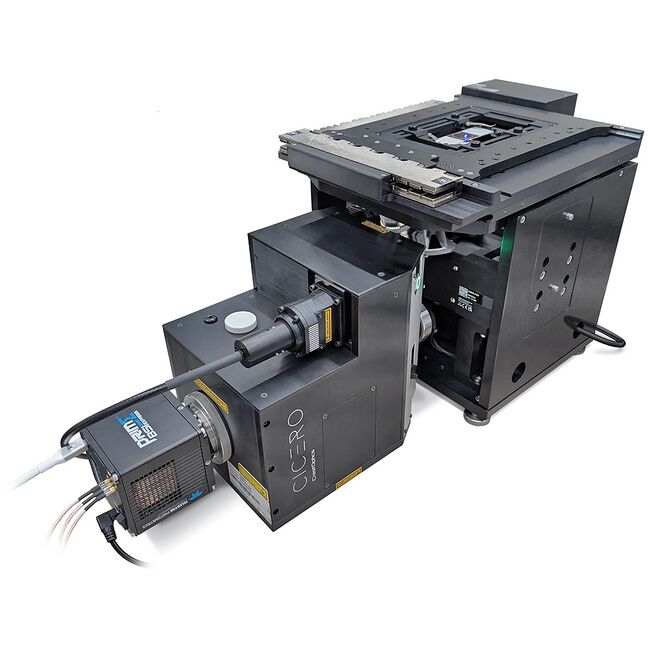
Figure 2. Zaber Nucleus Microscope and Crest Cicero spinning disc confocal
To streamline integration with larger confocal modules like the VTiSIM, Crest V3 and Yokogawa W1, Zaber customers have found that 25 mm pedestal mounts (Fig. 3) are useful. Pedestal mounts are available in a range of heights to match the microscope C-Mount height with the C-Mount height on the imaging module. Pedestal mounts and clamps also help to simplify setup by freeing you from the constraints of the fixed hole spacing of optical tables while still providing secure mounting.
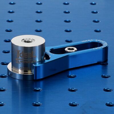
Figure 3. Pedestal mount and clamp (Courtesy of Ideal spectroscopy)
Full Support for Live Cell Imaging
Live cell imaging requires temperature, humidity, and frequently, CO2 control. Nucleus systems support three different methods of environmental control for live cells.
Option 1: Full Incubator-Ready Design
The small size of Nucleus inverted microscopes makes it possible to put the entire microscope system into an incubator at 37॰C. The humidity must be non-condensing. Not all third party imaging modules are incubator compatible.
Option 2: Cage Enclosure Integration
Incubated cage enclosures are available from Okolab (Fig. 4). These enclosures cover the XY stage and provide environmental control. They also block external light. Extended C-Mount ports are available for users of Nikon and Olympus objectives. This option provides additional clearance to make the integration of confocal and SIM units easier when using them in conjunction with the incubated enclosure. Cage incubators do not provide humidity and CO2 control.
Option 3: Stage-Top Incubator Support
Zaber ADR130 and ASR100 XY stages use the industry standard “K-Frame” aperture. These support stagetop enclosures from Ibidi, Okolab and Tokai Hit. Stage top incubators are ideal when humidity and CO2 control are required.
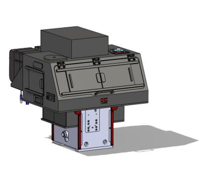
Figure 4. Okolab Bold microscope enclosure for Zaber Nucleus inverted microscope
Reliable, Long-Term Autofocus
In addition to incubation, live cell imaging typically requires autofocus to support long-duration imaging. Zaber’s HL04 microscope autofocus module provides reliable focus control for both scanning and timelapse imaging.
Simple, Powerful Software & Control
Hardware integration is only half the story. Once the system is set up, it must be controlled. Fortunately, software integration with Zaber is quick and easy.
Seamless 3rd-Party Software Integration
Native support for Zaber microscopes is included in Starlyte Nebula, Visiview, Inscoper, and μManager software packages. These software packages provide a GUI and support for fully automated image acquisition. To maximize throughput, Nebula, Visiview, Inscoper are recommended over μManager which can be significantly slower than more highly optimized packages. Depending on your needs and desired workflow once you have acquired your images, Starlyte Nebula, Inscoper and Visiview also provide increasingly advanced image processing capabilities. Zaber microscopes also support multi-dimensional image acquisition using our free Zaber Launcher software.
Advanced Custom Control with the ZML API
For advanced automation and integration with custom software tools and processes, Nucleus microscopes can be controlled with the Zaber Motion Library (ZML) API. ZML is available for Python, C++, C#, Java, JavaScript, Matlab, and Laview. The API is comprehensively documented and includes a large library of example code and application-specific whitepapers.
High-Speed Sync with 3rd-Party Hardware
Integration and control of third party hardware is also possible via IO triggering. The Nucleus microscope’s XY stage, focus stage, and LED lighting controller all have digital IO ports which can interface with external devices via TTL triggering. Triggers can be configured using our free Zaber Launcher software (Fig. 5).
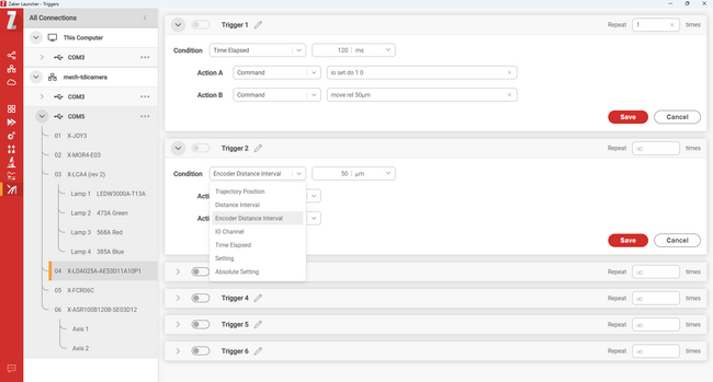
Figure 5. Configuring IO triggers in the free Zaber Launcher Software
Conclusion
Nucleus microscopes are an ideal platform for researchers who want to assemble their own high-resolution confocal or SIM imaging system. Zaber’s microscope platform makes this affordable and accessible, challenging the idea that such systems must be costly "black-box" purchases. Whether you're ready to start building or just have a few questions, contact our team for expert help.